

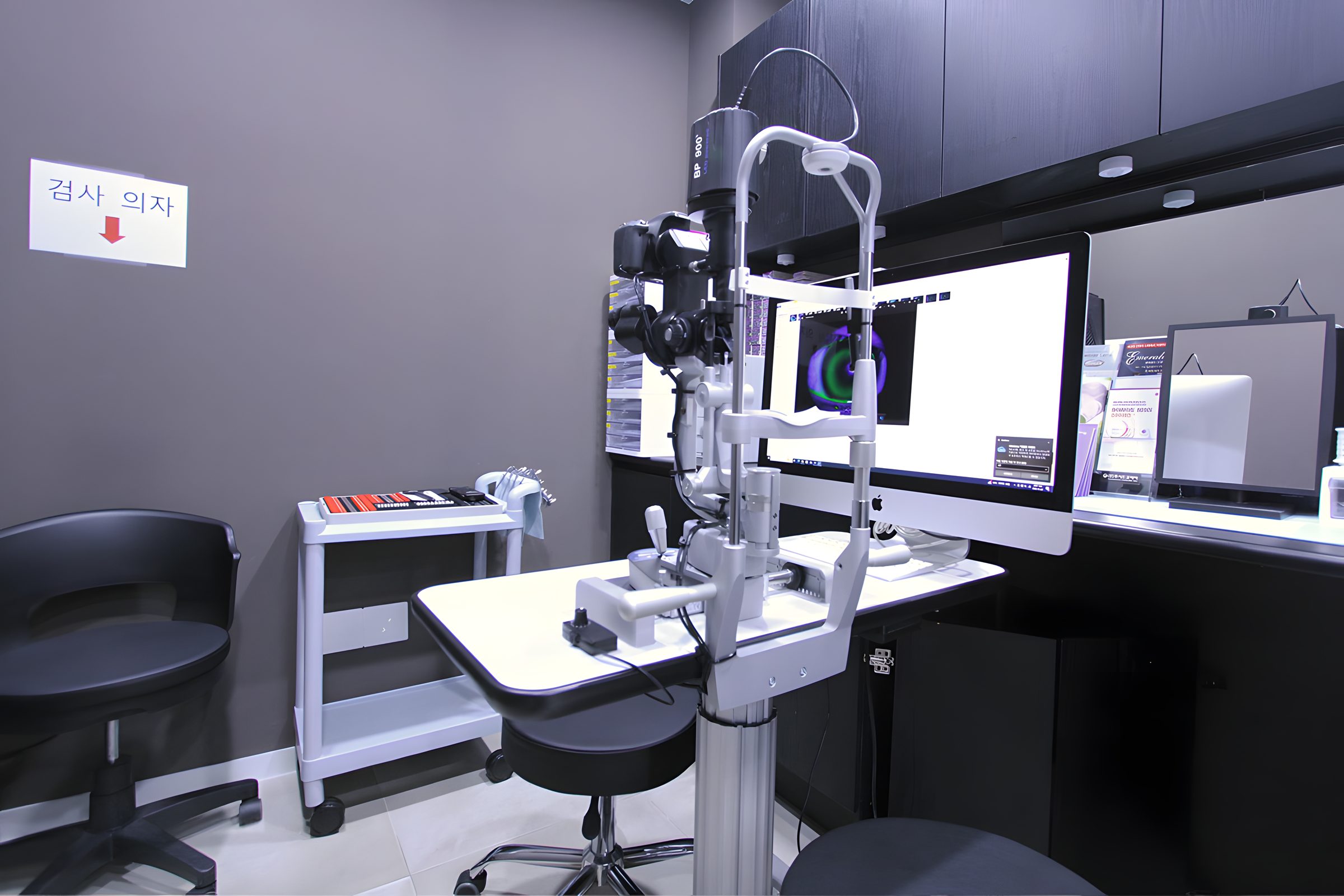
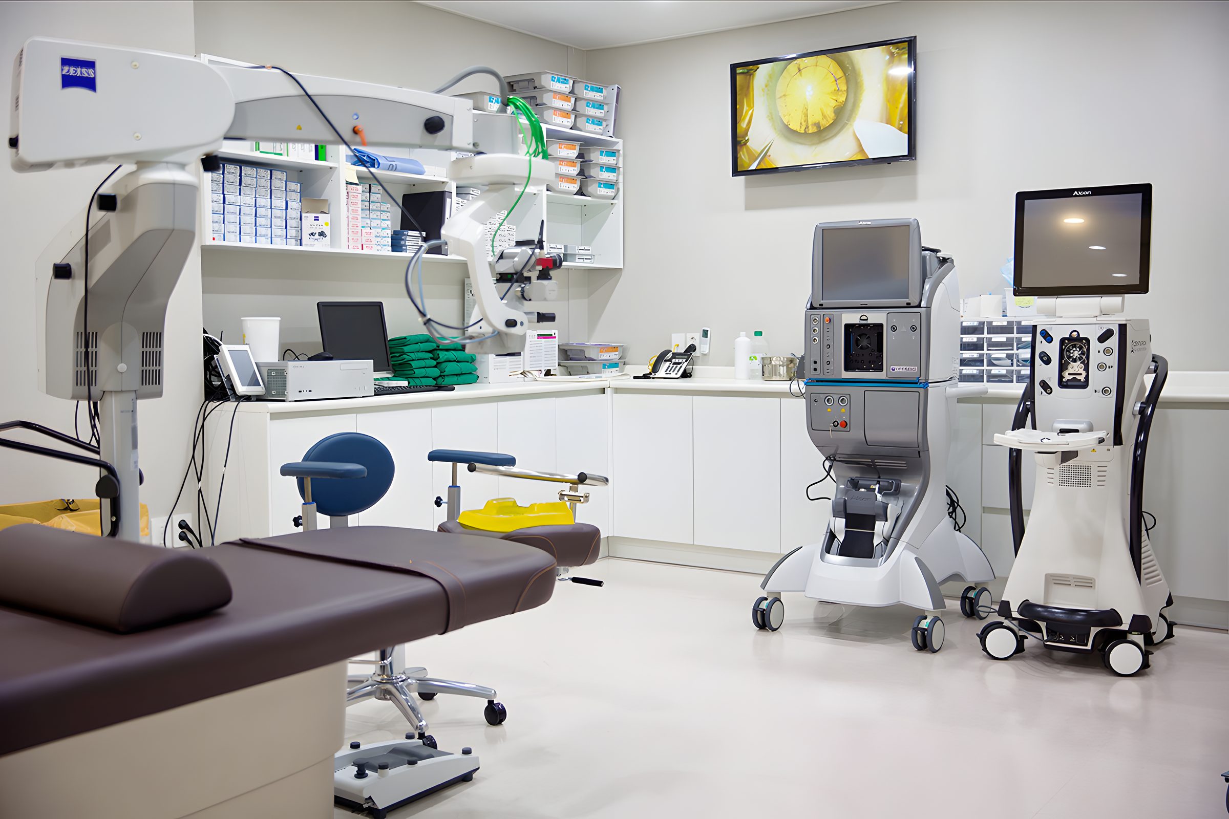
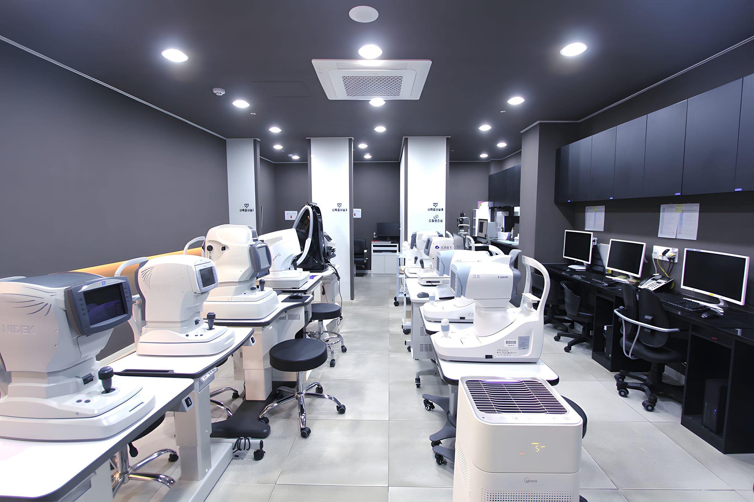

Jalboneun Seongmo Eye Clinic ZEISS® precision diagnostic equipment
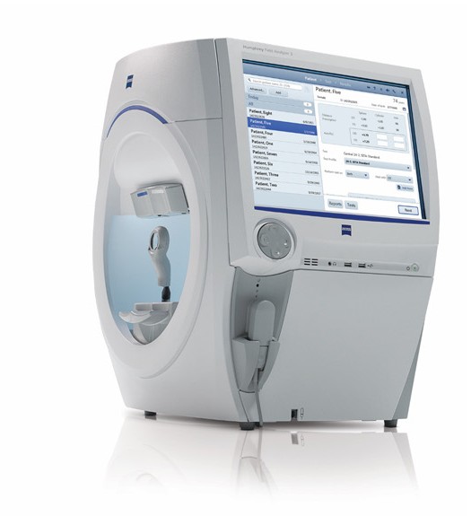
Our clinic can perform both static visual field testing and kinetic visual field testing (needed for disability certification and driver's license renewal).
Our clinic performs all visual field tests on the Humphrey® 840, a higher-end Zeiss perimeter model.
This is a higher-end model that can perform kinetic testing, a specialized version of visual field testing needed for disability certification and driver's license renewal.
Data from visual field testing can also be linked with Zeiss Forum so serial change can be assessed at a glance.
- DICOM Data Transfer
- DICOM MWL
- ZEISS FORUM
- HEYEX (HEIDELBERG ENGINEERING)
- PAS
Humphrey Field
Analyzer3 (HFA3)®
The Zeiss Humphrey Field Analyzer 3 (HFA3)® is known as an international standard among glaucoma specialists.
This perimeter can perform both static and kinetic visual field testing.
Precision is high, so fine changes in visual field defects can be measured accurately.
It therefore plays an important role in detecting visual field damage from glaucoma early and measuring how far it has progressed.
The test time is short, and patients can be tested comfortably.
- 01Static visual field test
Static visual field testing measures the degree of field loss by comparing the brightness of points shown on a computer screen. HFA3 can examine both the center and the periphery of the visual field with static testing.
- 02Kinetic visual field test
Kinetic visual field testing measures how far the visual field extends to the edge by moving a lighted target in front of the eye. Our 840 version can perform kinetic testing and print results needed for disability certification and driver's license renewal.
Glaucoma, for which early diagnosis is most important
Because there is no method that can cure glaucoma, finding it early and treating it is most important.
When glaucoma is found early and appropriate treatment is given, progression to blindness can be prevented.
The core of glaucoma care is early detection and steady treatment.
A glaucoma specialist sees patients across glaucoma, from refractory neovascular glaucoma to pediatric glaucoma.
Comparison of glaucoma diagnostic accuracy between built-in RNFL thickness indices on optical coherence tomography and newly calculated indices — Kang KD, Park CK (Department of Ophthalmology, College of Medicine, The Catholic University of Korea), J Korean Ophthalmol Soc Vol. 47 No. 2 2006
Purpose: To determine which indices from optic-disc analysis and RNFL thickness analysis by optical coherence tomography are most useful for diagnosing glaucoma, and to compare them with built-in normative indices (99%, 95% values). Methods: 499 eyes of 284 people were classified as normal, glaucoma-suspect, or glaucoma; optic-disc analysis and RNFL thickness analysis were performed and results were reviewed retrospectively. Results: There were 134 normal eyes, 128 glaucoma-suspect eyes, and 237 glaucoma eyes; all six RNFL thickness indices differed significantly between the glaucoma and non-glaucoma groups. Among indices, average RNFL thickness showed the highest correlation with visual field defects and the highest discriminant power at a cutoff value of 92.39 mm², followed by inferior average RNFL thickness. A discriminant formula combining age, Horizontal Integrated Rim Area, and average RNFL thickness had higher discriminant power than each alone, with accuracy 76.2%, sensitivity 73.1%, and specificity 73.1%. Conclusion: Average RNFL thickness then inferior average RNFL thickness were useful for diagnosing glaucoma, and the new discriminant formula was better in sensitivity and accuracy than built-in indices and is useful for glaucoma diagnosis. (J Korean Ophthalmol Soc 47(2):243-252, 2006)
Discrimination of glaucoma in children using indices obtained by optical coherence tomography — Kang KD, Aman Shah B. Abdul Majid, Kim Yeondeok, Gwak Jihyeon, Im Hyebin, J Korean Ophthalmol Soc 2009 Vol. 50 No. 6
Purpose: To determine which OCT-derived indices are useful for diagnosing glaucoma in children. Methods: Eighty-four children aged 6 to 18 years (127 eyes) who visited the ophthalmology department from March 2006 to February 2008 underwent ophthalmic examination and retinal nerve fiber layer (RNFL) thickness analysis by OCT. Subjects were classified as normal, glaucoma suspect, or glaucoma and reviewed retrospectively. Results: Fifty-five eyes were normal, 27 glaucoma suspect, and 45 glaucoma. The index with the highest correlation and sensitivity for glaucoma was average RNFL thickness, followed by inferior and superior RNFL thickness. A discriminant formula combining superior, inferior, and average RNFL thickness had higher discriminant power than each index alone, with sensitivity 78% and specificity 68.6%. Conclusion: Among OCT indices, average RNFL thickness then inferior RNFL thickness were useful for diagnosing glaucoma, and a new discriminant formula with high sensitivity and specificity is useful for glaucoma diagnosis. (J Korean Ophthalmol Soc 2009:50(6):887-892)
A case of bleb dysesthesia treated with aqueous drainage-device implantation (case report) — Kang KD, Kim Yeondeok, Aman Shah B. Abdul Majid, Gwak Jihyeon, John Salmon, Im Hyebin, J Korean Ophthalmol Soc 2010 Vol. 51 No. 5
Purpose: To report effective treatment of bleb dysesthesia with aqueous drainage-device implantation. Case summary: A 37-year-old woman who had undergone trabeculectomy in the left eye for persistent intraocular pressure elevation despite medical therapy presented with intermittent foreign-body sensation and pain in the same eye. No pressure elevation was observed. Slit-lamp examination showed punctate corneal epithelial damage below the bleb; bleb dysesthesia was diagnosed and artificial tears were prescribed. Symptoms persisted, so a bandage contact lens, bleb compression suture, and bleb revision were performed in sequence, still without improvement. Aqueous drainage-device implantation was then performed, and bleb excision 3 months later yielded a very satisfactory improvement in symptoms. Conclusion: Aqueous drainage-device implantation can be an effective alternative for dysesthesia after trabeculectomy, and bleb dysesthesia should be explained in detail to the patient before trabeculectomy. (J Korean Ophthalmol Soc 2010:51(5):790-793)
Comparison of cost and distribution by primary treatment in patients with primary open-angle glaucoma — Gwak Dongyun, Gwak Jihyeon, Kim Yeondeok, Im Hyebin, Baek Hyejeong, Park Chijun, Aman Shah B. Abdul Majid, Kang KD, J Korean Ophthalmol Soc 2010 Vol. 51 No. 6
Purpose: To examine, in the Korean setting, the cost over time and the distribution of patients when medication, laser, or surgery is chosen as initial glaucoma treatment. Methods: An independent Markov process was built for each of the three initial treatments to construct a Markov model and show the probabilistic distribution of states over time. One stage was defined as 1 year, and costs over 5, 10, and 20 years were obtained by Monte Carlo simulation. Results: At 5 years, choosing laser as initial treatment had the lowest cost; at 10 years, choosing surgery had the lowest cost. At 10 years, surgery as initial treatment is more economical than medication if the surgical success rate is 30.1% or higher; laser as initial treatment is more economical than medication if the laser success rate is 16.3% or higher. Annual drug cost would need to fall to about 55,000 won or 60,000 won or less to be more economical than surgery and laser, respectively. Conclusion: Laser trabeculoplasty has favorable cost-effectiveness over a short period (5 years), and surgery has favorable cost-effectiveness over a long period (10 years or more). (J Korean Ophthalmol Soc 2010:51(6):865-874)